Establishment of a fluorescent in situ hybridization assay for imaging hepatitis B virus nucleic acids in cell culture models
Xiaonan Zhang (张小楠), Lei Yue(岳蕾), Zhanqing Zhang(张占卿), and Zhenghong Yuan(袁正宏)
Shanghai Public Health Clinical Center, Key Laboratory of Medical Molecular Virology, Shanghai Medical College, Fudan University
While chronic hepatitis B remains a global public health problem, the detailed spatiotemporal dynamics of the key molecular events leading to the multiplication and egress of hepatitis B virus is still largely unclear. Previously, we developed a chromogenic in situ hybridization (ISH) assay for detection of HBV RNA, DNA and cccDNA in clinical liver biopsies. Herein, we report the establishment of a fluorescent in situ hybridization (FISH) method for visualization of HBV RNA, HBV core particle DNA and intranuclear DNA in a tetracycline inducible HBV replication system (HepAD38) and a de novo infection system (HepG2-NTCP). Utilizing 3D-STORM, we were able to obtain images of HBV RNA and DNA with improved spatial resolution allowing in-depth analysis of key virological events within complex subcellular compartments. Taken together, these techniques should facilitate a deeper understanding of the molecular behavior of HBV life cycle and shed new light on the intricate mechanisms of virus-host interaction.While chronic hepatitis B remains a global public health problem, the detailed spatiotemporal dynamics of the key molecular events leading to the multiplication and egress of hepatitis B virus is still largely unclear. Previously, we developed a chromogenic in situ hybridization (ISH) assay for detection of HBV RNA, DNA and cccDNA in clinical liver biopsies. Herein, we report the establishment of a fluorescent in situ hybridization (FISH) method for visualization of HBV RNA, HBV core particle DNA and intranuclear DNA in a tetracycline inducible HBV replication system (HepAD38) and a de novo infection system (HepG2-NTCP). Utilizing 3D-STORM, we were able to obtain images of HBV RNA and DNA with improved spatial resolution allowing in-depth analysis of key virological events within complex subcellular compartments. Taken together, these techniques should facilitate a deeper understanding of the molecular behavior of HBV life cycle and shed new light on the intricate mechanisms of virus-host interaction.
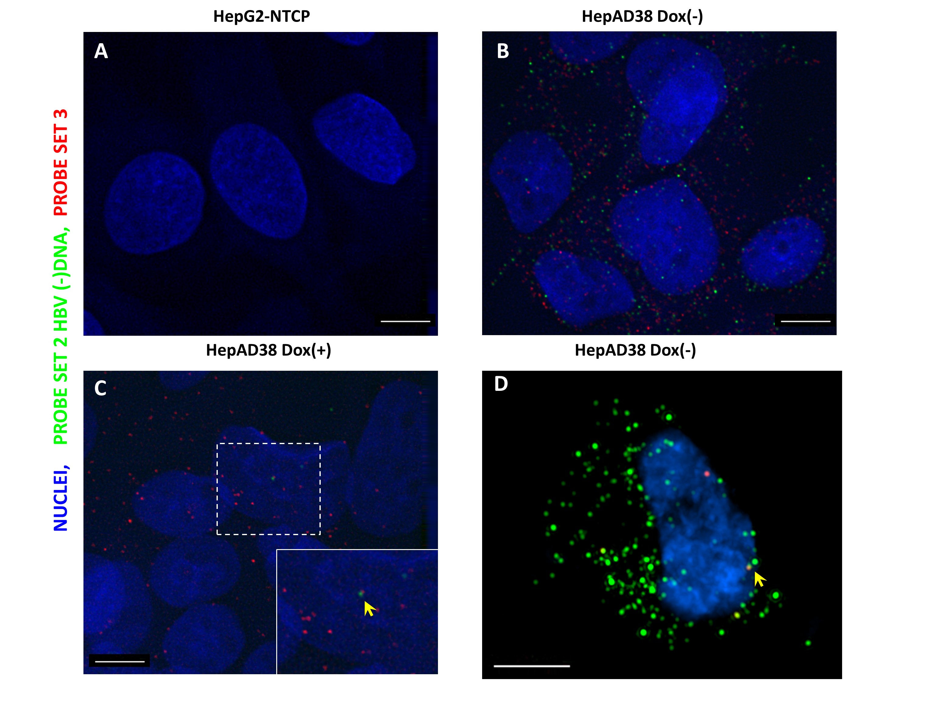
Fluorescent in situ hybridization of HBV RNA and DNA in a hepatoma cell line supporting HBV replication. HepG2-NTCP (A) and HepAD38 (B-D) cells, that are either maintained in doxycycline supplemented medium (C) or doxycycline-free medium, were fixed and permeabilized followed by hybridization with Probe set 2 and probe set 3. After branch DNA amplification, fluorophore conjugated label probe was applied and images were acquired in a Deltavision epifluorescence microscope (A-C) and Nikon 3D-SIM microscope (D).

Nikon 3D-SIM (Structural illumination) imaging of HBV nucleic acids in HepAD38 cells
Image Collections
Type of Staining
- HBV RNA(red)-HBV DNA(-strand,green)
- HBV DNA (-strand, green)