Spatial and functional links between cellular virological state and progression of liver fibrosis in chronic hepatitis B
Xiaonan Zhang (张小楠), Danping Liu (刘丹萍), Wei Lu(陆伟), Ye Zheng(郑叶), Min Wu(邬敏),Jiahui Ding(丁家惠),Mingzhu Xu(徐明珠), Xiaohui Zhou (周晓辉),Yanling Feng(冯艳玲), Zhanqing Zhang(张占卿), and Zhenghong Yuan(袁正宏)
Shanghai Public Health Clinical Center, Key Laboratory of Medical Molecular Virology, Shanghai Medical College, Fudan University
https://www.biorxiv.org/content/10.1101/2020.01.13.904201v1
Chronic
Hepatitis B Virus (HBV) infection is strongly associated with the progression
of liver fibrosis, cirrhosis and hepatocellular carcinoma. Despite intensive
study, the detailed mechanisms leading to HBV induced liver disease have not
been fully elucidated. Previously, we reported a mosaic distribution of viral
antigens and nucleic acids at single-cell level in liver tissues of chronic
hepatitis B (CHB) patients and proposed a ‘three-stage model’ of HBV infection
in vivo. Here, we explored whether the different stages at cellular level is
functionally linked with fibrogenesis. Strikingly, we observed a tight spatial
relationship between the invasion of collagen fibers and transitions from
S-rich to DNA-rich stage. While S-rich cells mainly localized within minimally
fibrotic tissue, DNA-rich cells were often closely surrounded by a milieu of
stiffened extracellular matrix (ECM). cDNA microarray and subsequent validation
analyses revealed that S-rich cells manifested elevated ribosomal proteins and
oxidative phosphorylation genes in a disease phase-dependent manner. On the
other hand, DNA-rich cells exhibited gradually deteriorated expression of
hepatocyte-specific antigen and transcriptional regulator in parallel with the
progression of hepatic fibrosis. Finally, during fibrogenesis, inflammatory
genes such as IP-10 were found to be expressed in both portal infiltrated cells
and surrounding parenchymal cells which resulted in suppressed antigen
expression.
Based on the observations in this study, we propose an updated model of HBV life-cycle in a histological context, in which inflammation and fibrogenesis are incorporated (See the figure below ). In immune quiescent phase of the CHB infection (equivalent to fibrosis stage 0-1), the majority of the HBV infected cells exhibited an S-rich state. The infected cells harbor high level of viral antigens such as HBsAg and possibly HBx which boost some of the essential cellular activities such as protein synthesis and energy conversion. Spontaneous transition into DNA-rich state is initiated due to the loss of immune-quiescence and subsequent activation of HSCs and deposition of collagen fibers (equivalent to fibrosis stage 2-3). The infiltration of immune cells and resulting secretion of inflammatory mediators trigger hepatocellular inflammation gene expression and suppress the expression of viral antigens and might also contribute to S-rich to DNA-rich transition. The stiffened ECM milieu and associated inflammation inhibited the major hepatocellular functions by down-regulating key transcription factors such as HNF4a. Further accumulation of rigid ECM (pseudolobule formation) around HBV infected hepatocytes eventually leads to the deterioration of these cells, part of which are finally immersed into the fibrotic tissue (equivalent to fibrosis stage 4). As a result of inflammation and advanced fibrosis, a significant number of HBV positive cells convert to the latent stage with minimal viral expression and replication.
Taken together, we propose that liver inflammation and accompanying fibrogenesis is spatially and functionally linked with the transition of virological stages at cellular level. These transitions occur possibly due to an altered hepatocyte transcription profile in response to a transformed ECM environment. The collective viral and host activities shape the histological alterations and progression of liver disease during CHB infection.
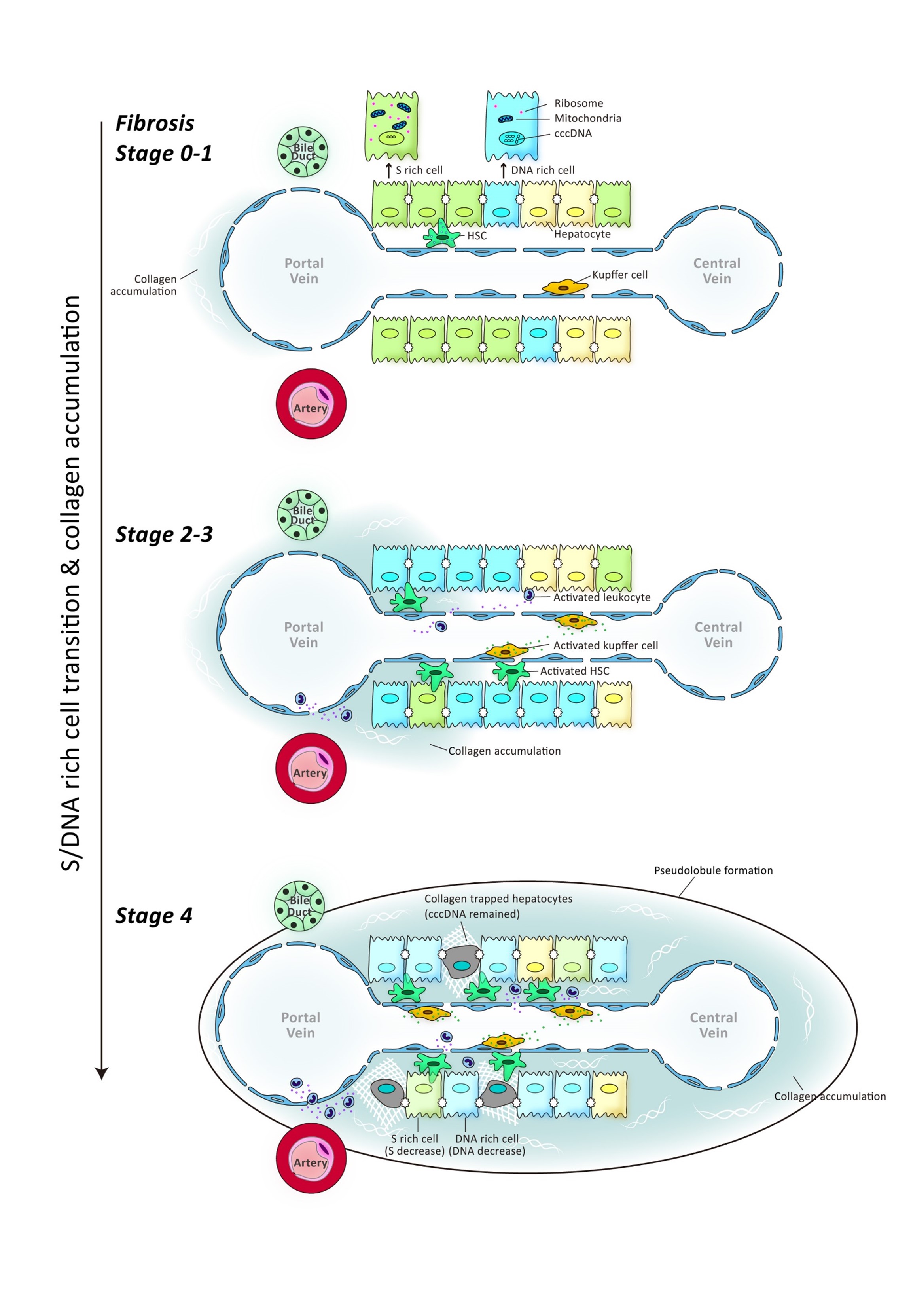
Schematic illustration of the updated model of
HBV life-cycle in the histological context.
Image Collections
Type of Staining
- HBVDNA-S-Sirius Red
- HBVDNA-HNF4a-Sirius red
- HBVDNA(blue)-Hep-Par1(red)
- IP10(blue)-HBsAg(brown)-hematoyxlin
- IP10(blue)-HBsAg(brown)-Sirius red
- IP10(blue)-HBVDNA(yellow)-HBsAg(red)
- HBsAg(red)-CollagenI(green) IFA
- HBVDNA(red)-CollagenI (green) IFA